


Article / Research Article
Orthotics and Prosthetics Research Clinic, Tehran, Iran.
Dr. Sedegheh Malek Mohammadi,
Orthotics and Prosthetics Research Clinic,
Tehran,
Iran.
28 February 2025 ; 15 March 2025
Plantar fascia connects the calcaneus bone to the plantar aspect of the toes and attaches to the soft tissues and bones (Aquino & Payne, 1999). Plantar fascia supports the medial longitudinal arch of the foot (Caravaggi et al., 2010). It has important role in dynamic and static positions of the foot through maintaining the height of the medial longitudinal arch (Gefen, 2002). It is believed functional role of the plantar fascia is dependent on its mechanical properties (Wearing et al., 2006).
The objective of this research is better understanding the structure and properties of the plantar fascia.
Method
- To establish what are known about the characteristics of the plantar fascia.
- To locate specific property of the plantar fascia.
The properties information of the plantar fascia can be source of collecting data according to the biological properties of the body in living individual, as close as possible.
Keywords: kinesiology; Biomechanics; Plantar fascia thickness; Vertical load.
Plantar fascia consists of three bands and is divided in different depths and planes and attaches to the medial and lateral intermuscular septa. It attaches to the soft tissues and bones. (flexor digitorum brevis, abductor hallucis, calcaneus, base of the fifth metatarsal, skin, deep transverse metatarsal ligament, medial and lateral intermuscular septa) (Aquino & Payne, 1999). It is assumed plantar fascia is supporting medial longitudinal arch of the foot, also (Gefen, 2002). Plantar fascia elongates in loading and stores energy but plantar fascia has a limited ability to elongate (Bartold 2004). It is believed plantar fascia like ligaments has viscoelastic properties but there are histological differences between plantar fascia and ligaments (Wearing et al., 2006).
![]() Figure 1: Body weight transmission
Figure 1: Body weight transmission
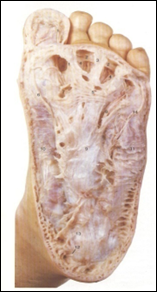 Figure 2: Plantar fascia
The body weight is applied on the foot through the shank and ankle joint (Gefen, 2003). On the attached bony segments of the plantar fascia (calcaneus and head of the first metatarsal bone), the vector of the applied load may be divided into two vectors. Although the vertical vector may be counteracted with the opposite vertical vector of the ground reaction force, (Bolgla & Malone, 2004) but the plantar fascia may be elongated with two horizontal vectors in opposite directions. That’s why it has been decided properties of the plantar fascia are studied biologically.
The plantar fascia is a thick, multilayered and inelastic band of fibrous tissue that extends along the plantar surface of the foot (Jeswani et al., 2009). Plantar fascia includes of collagen and elastic fibers. Elastic fibers are shaped as strands and networks with different thicknesses. The elastic fibers in compare with the collagen fibers have lower modulus of elasticity. Plantar fascia is composed of densely compacted collagen fibers that most of them are originated longitudinally and some of them are directed transversely and obliquely (La Porta & La Fata, 2005). Plantar fascia originates from the medial process of the calcaneal tuberosity (Wearing et al. 2006) and proximal to the origin of the flexor digitorum brevis (La Porta & La Fata, 2005). The medial band of the plantar fascia is thin and non-existent at its proximal level; on the contrary, the lateral band is thick and developed but it is completely absent in 12% of people. The central aponeurotic band is the major component of the plantar fascia both structurally and functionally. Its origin is independent of the tendo-achilles in adults. At the level of mid-tarsal, central band is divided into five branches and proximal to the metatarsal heads, each of them is divided into superficial and deep tracts (Wearing et al., 2006). The medial and the lateral bands cover the intrinsic musculature of hallux and fifth digit. The central band is the most tensile band and overlies the short and long digital musculature. There are longitudinal vertical fibers between the medial and central and between the central and lateral bands that form the intermuscular septa; besides, the branches of plantar vessels and nerves are sent to the sole of the foot through them (La Porta & La Fata, 2005).
The plantar fascia is innervated by two nerves which arise from the posterior tibial nerve. The first is the medial calcaneal nerve (MCN) which is the last branch of the posterior tibial nerve before dividing into the medial and lateral plantar nerves (MPN and LPN). Its location is between the abductor hallucis and superficial fascia. The second one is the first branch of the lateral plantar nerve. It passes along the medial border of the calcaneus before passing between abductor hallucis and quadrates platae muscles, then it passes through a soft tissue tunnel between abductor hallucis and flexor digitorum brevis muscles before reaching the plantar fascia (McNally et al., 2010). Nerves pass from the internal side of the equivalent arteries and are parallel with them. The braches of plantar nerves and vessels are sent to the sole of the foot through intermuscular septa (La Porta & La Fata, 2005).
 (Alshami et al., 2008)
Figure 3: Innervations and circulation of the plantar fascia
Thickness of the plantar fascia is different along the length of the plantar fascia. It is modified under loading, also (Garcia et al., 2008). Thickness of the plantar fascia is parameter to identify the location along the length of the plantar fascia (Huerta, 2007). The mid substance of the plantar fascia contains fibroblast cells within wavy collagen fibers (extra cellular matrix) (La Porta & La Fata, 2005; Wearing et al., 2006) plantar fascia elongates under loading. (Bartold, 2004) fibroblast cells reshape (Grinnell, 2008) and wavy collagen fibers are straightened under loading (Aquino & Payne, 1999). They back again during unloading (Langevin et al., 2009). With regard to the changing of the thickness along the length of the plantar fascia, it can be either the address of each location of the plantar fascia or a biomechanical variable to understand the concentration of the amount of the identified load applied on the plantar fascia (Wearing et al., 2006; Garcia et al., 2008).
 Figure 4: Plantar fascia properties
There are different characteristics between the plantar fascia and fascia tissue in the body.
Plantar fascia has specific properties in the body of each person.
In the body of living individual, plantar fascia properties of both feet are not equal.
When plantar fascia thickness is put under vertical load, dynamic and static position of the foot and supporting of the height of the medial longitudinal arch is possible.
- Aquino, A., & Payne, C. (1999). Function of the plantar fascia. The foot, 9(2), 73-782. DOI: https://doi.org/10.1054/foot.1999.0520
- Caravaggi, P., Pataky, T., Günther, M., Savage, R., & Crompton, R. (2010). Dynamics of longitudinal arch support in relation to walking speed: contributiFon of the plantar aponeurosis. Journal of Anatomy, 217(3), 254-261. DOI: https://doi.org/10.1111/j.1469-7580.2010.01261.x
- Gefen, A. (2002). Stress analysis of the standing foot following surgical plantar fascia release. Journal of Biomechanics, 35(5), 629-637.
DOI: https://doi.org/10.1016/s0021-9290(01)00242-1 - Wearing, S. C., Smeathers, J. E., Urry, S. R., Hennig, E. M., & Hills, A. P. (2006). The pathomechanics of plantar fasciitis. Sports Medicine, 36(7), 585-611. DOI:https://doi.org/10.2165/00007256-200636070-00004
- Bartold, S. J. (2004). The plantar fascia as a source of pain-biomechanics, presentation and treatment. Jurnal of bodywork and movement therapies, 8(3), 214-226. DOI: https://doi.org/10.1016/S1360-8592(03)00087-1
- Gefen, A. (2003). The in vivo elastic properties of the plantar fascia during the contact phase of walking. Foot & Ankle International, 24(3), 238-244.
DOI: https://doi.org/10.1177/107110070302400307 - Bolgla, L. A., & Malone, T. R. (2004). Plantar fasciitis and the windlass mechanism: A biomechanical link to clinical practice. Journal of Athletic Training, 39(1), 77-82. https://pmc.ncbi.nlm.nih.gov/articles/PMC385265/
- Jeswani, T., Morlese, J., & McNally, E. G. (2009). Getting to the heel of the problem: plantar fascia lesions. Clinical Radiology, 64(9), 931-939.
DOI: https://doi.org/10.1016/j.crad.2009.02.020 - La Porta, G. A., & La Fata, P. C. (2005). Pathologic conditions of the plantar fascia. Clin Podiatr Med Surg, 22(1), 1-9.
DOI: https://doi.org/10.1016/j.cpm.2004.08.001 - McNally, E. G., & Shetty, S. (2010). Plantar fascia: Imaging Diagnosis and Guided Treatment. Semin Musculoskeletal Radiol, 14(3), 334-343.
DOI: https://doi.org/10.1055/s-0030-1254522 - Garcia, C. A., Hoffman, S. L., Hastings, M. K., Klaesner, J. W., & Mueller, M. J. (2008). Effect of metatarsal Phalangeal joint extension on plantar soft tissue stiffness and thickness. Foot (Edinb), 18(2), 61-7. DOI: https://doi.org/10.1016/j.foot.2007.12.002
- Huerta, J. P., & Alarcón García, J. M, (2007). Effect of gender, age and anthropometric variables on plantar fascia thickness at different locations in asymptomatic subjects. European Jurnal of Radiology, 62(3), 449-453. DOI: https://doi.org/10.1016/j.ejrad.2007.01.002
- Grinnell, F. (2008). Fibroblast mechanics in three-dimensional collagen matrices. Journal of Bodywork Movement, 12(3), 191-193.
DOI: https://doi.org/10.1016/j.jbmt.2008.03.005 - Langevin, H. M., & Huijing, P. A. (2009). Huijing, Communication About Fascia: History, Pitfalls, and Recommendation. Int J Ther Massage Bodywork, 2(4), 3–8. DOI: https://doi.org/10.3822/ijtmb.v2i4.63.