

Article / Case Report
Department of Internal Medicine, Catholic Health Initiatives (CHI) St. Vincent, Hot Springs, Arkansas, USA.
Sameer Kapadia, MD,
Department of Internal Medicine Catholic Health Initiatives (CHI) St. Vincent Hot Springs 300 Werner Avenue Hot Springs, AR 71913, USA.
25 September 2026 ; 1 October 2026 ; 15 October 2026
Citation: Kapadia, S., Howard, M., & Pabona, J. M. P. (2026). Large Jejunal Villous Adenoma Mimicking Malignancy in a 90-Year-Old Man. J Medical Case Repo 8(5):1-4.
Small-bowel neoplasms are uncommon and comprise a heterogeneous group of benign and malignant lesions. Small-bowel cancers account for only a small proportion of gastrointestinal malignancies, with adenocarcinoma, neuroendocrine tumors, lymphoma, and gastrointestinal stromal tumors representing important malignant entities (Raghav & Overman, 2013). Despite advances in cross-sectional imaging, capsule endoscopy, and device-assisted enteroscopy, diagnosis of small-bowel tumors can remain challenging because of their relatively low incidence, nonspecific clinical presentation, and anatomic location (Cheung et al., 2016; Islam et al., 2014; Kim, 2020).
Adenomas of the small intestine are also uncommon. Histologically, they share features with colorectal adenomas and are recognized as precursor lesions in the adenoma-carcinoma sequence of the small bowel (Perzin & Bridge, 1981). Villous tumors occur predominantly in the proximal small intestine, particularly the duodenum, while true villous adenomas arising in the jejunum are distinctly rare (Delevett & Cuello, 1975; Keeley & Gottlieb, 1969; Mir-Madjlessi et al., 1973; Wiseman et al., 2007). When sufficiently large, these lesions may cause gastrointestinal bleeding, obstruction, or intussusception.
We present a case of a large proximal jejunal villous adenoma associated with intussusception and small-bowel obstruction in a 90-year-old man. His presentation was notable for substantial unintentional weight loss and findings concerning for malignancy, although final pathology demonstrated a mucosa-confined villous adenoma with low-grade dysplasia and no invasive carcinoma.
A 90-year-old man with a history of atrial fibrillation, sick sinus syndrome, coronary artery disease, chronic systolic heart failure, hypertension, hyperlipidemia, and gastroesophageal reflux disease presented for surgical evaluation of an enlarging right inguinal hernia. The patient reported that the right groin bulge had progressively enlarged over approximately six months and was associated with discomfort, particularly with sitting and standing. The bulge became more prominent with coughing, straining, and lifting.
During the same six-month period, he developed early satiety, intermittent emesis, decreased energy, and approximately 60 lb of unintentional weight loss. He denied night sweats, hematochezia, or melena. He had not undergone endoscopic evaluation.
Initial laboratory evaluation demonstrated a hemoglobin of 11.9 g/dL, white blood cell count of 9.4 × 10³/µL, platelet count of 109 × 10³/µL, sodium of 131 mmol/L, potassium of 5.3 mmol/L, bicarbonate of 20 mmol/L, blood urea nitrogen of 25 mg/dL, and creatinine of 1.2 mg/dL.
Because of his persistent gastrointestinal symptoms and marked unintentional weight loss, prior abdominal imaging was reviewed by the surgical team. Review of a CT abdomen and pelvis obtained on October 11, 2025 demonstrated proximal jejunal intussusception. A repeat CT obtained with oral contrast on October 27, 2025 demonstrated a suspected proximal jejunal transition point with associated luminal narrowing (Figure 1). This region corresponded to the proximal jejunal segment subsequently found intraoperatively to contain the obstructing intraluminal mass. A water-soluble contrast enema performed on October 31, 2025 was without evidence of a significant colonic lesion. Review of an earlier CT from June 24, 2021 showed no evidence of a comparable small-bowel lesion.
The imaging findings, progressive gastrointestinal symptoms, and 60-lb unintentional weight loss raised concern for an underlying small-bowel neoplasm. The differential diagnosis included adenocarcinoma, gastrointestinal stromal tumor, neuroendocrine tumor, lymphoma, and an adenomatous lesion.
The patient underwent operative exploration in conjunction with repair of the right inguinal hernia. During the hernia repair, the hernia sac was opened and the bowel was reduced. The bowel was viable, and moderate ascites was noted. The hernia was subsequently repaired with mesh.
An upper midline laparotomy was then performed. The small bowel was examined beginning at the ligament of Treitz, and a very proximal jejunal mass associated with intussusception was identified. The bowel was divided proximal and distal to the lesion, and the involved segment was resected. Because of the proximity of the lesion to the ligament of Treitz, reconstruction was performed with a handsewn end-to-end anastomosis after mobilization of the distal duodenum.
The resected segment measured 6.8 cm in length. When opened, it contained a circumferential, nearly obstructing polypoid lesion that was concerning for an advanced adenoma or malignancy. Gross pathologic examination demonstrated a 6.4 × 5.3 × 0.8 cm red-tan polypoid mass. The lesion was 1.2 cm from the closest mesenteric margin, 1.8 cm from the closest stapled margin, and 2.6 cm from the opposite stapled margin.
Histopathologic examination demonstrated a villous adenoma with low-grade dysplasia (Figure 2). The lesion was confined to the mucosa without involvement of the muscularis propria. There was no high-grade dysplasia or invasive carcinoma. All resection margins were negative for adenoma. Minimal surrounding fibroadipose tissue was submitted, and no lymph nodes were identified. Examination of the right inguinal hernia sac demonstrated chronic inflammation.
The postoperative course was complicated by a prolonged ileus, which subsequently resolved. Bowel function returned, his diet was advanced, and his pain was adequately controlled. He was discharged to rehabilitation on postoperative day 7.
Villous adenomas of the jejunum are rare, with relatively few cases described in the literature. Keeley and Gottlieb (1969) reported a large circumferential benign villous adenoma of the jejunum associated with intermittent bowel obstruction, hemorrhage, and intussusception, demonstrating that even a histologically benign villous lesion can produce significant mechanical complications. Mir-Madjlessi et al. (1973) subsequently reported villous tumors involving the duodenum and jejunum and reviewed the limited literature available at that time. Delevett and Cuello (1975) described another true villous adenoma of the jejunum. Similar to the present case, their lesion was benign and presented with intermittent intussusception.
More recently, Wiseman et al. (2007) described a proximal jejunal adenoma presenting with gastrointestinal bleeding. Subedi et al. (2022) reported a small-bowel adenoma producing jejunojejunal intussusception in a young adult. Together, these reports demonstrate the variable presentation of jejunal adenomas and their ability to produce bleeding, obstruction, or intussusception despite benign histology.
Adult intussusception differs from its pediatric counterpart because an underlying structural lesion is frequently present. Identifiable lead points include benign tumors, malignant neoplasms, inflammatory lesions, Meckel diverticula, and postoperative abnormalities (Marinis et al., 2009).
More contemporary evidence suggests that both the presence and malignant potential of a lead point vary according to the location of the intussusception. A systematic review of 37 studies involving 2,330 adult patients identified a pathologic lead point in 58% of cases (Heersche et al., 2025). Malignancy was identified in 9% of enteric intussusceptions compared with 68% of colocolic intussusceptions (Heersche et al., 2025). Thus, although malignancy remains an important consideration in adults with intussusception, benign structural lesions are also important causes of small-bowel intussusception.
In the present case, the 6.4-cm villous adenoma likely served as the structural lead point for the proximal jejunal intussusception. The lesion was also circumferential and nearly obstructed the bowel lumen, providing an additional mechanical explanation for the patient’s symptoms. Together, the intussusception and marked luminal narrowing likely contributed to his progressive early satiety and intermittent emesis.
The histopathologic findings are particularly notable given the size and clinical presentation of the lesion. Small-bowel adenomas are recognized precursor lesions for adenocarcinoma. In a clinicopathologic review of 51 small-bowel tumors containing adenomatous epithelium, Perzin and Bridge (1981) demonstrated an association between small-bowel adenomas and carcinoma and found that villous morphology and increasing lesion size were associated with a greater likelihood of carcinoma. The adenoma-carcinoma sequence is also recognized in the pathogenesis of small-bowel adenocarcinoma (Raghav & Overman, 2013). Despite its large size, villous morphology, and concerning clinical presentation, the lesion in our patient remained confined to the mucosa and demonstrated only low-grade dysplasia, without high-grade dysplasia or invasive carcinoma.
The patient’s presentation was concerning for malignancy because of his advanced age, substantial unintentional weight loss, and large small-bowel mass. However, benign small-bowel tumors can produce similar symptoms when they cause luminal narrowing, obstruction, bleeding, or intussusception. Modern diagnostic approaches to suspected small-bowel tumors include cross-sectional imaging, capsule endoscopy, and device-assisted enteroscopy, with the choice of modality depending on the clinical presentation and suspected lesion (Cheung et al., 2016; Fantasia et al., 2024; Islam et al., 2014; Kim, 2020). In this case, the presence of a large proximal lesion associated with intussusception and mechanical narrowing, together with the inability to exclude malignancy, favored operative management. Surgical resection provided both definitive diagnosis and treatment.
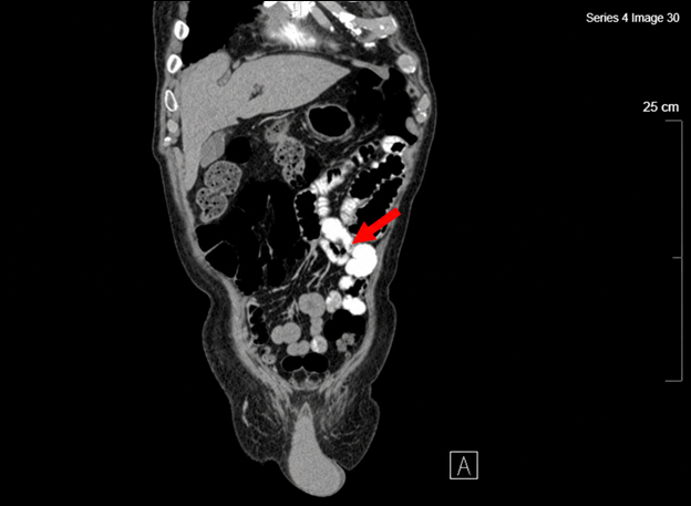 Figure 1: CT abdomen and pelvis with oral contrast obtained October 27, 2025 demonstrating a suspected proximal jejunal transition point with associated luminal narrowing (arrow). At operative exploration, this proximal jejunal segment was found to contain an obstructing intraluminal mass, subsequently confirmed on histopathology as a villous adenoma with low-grade dysplasia.
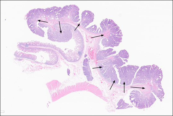 Figure 2: Histopathologic examination of the resected jejunal villous adenoma demonstrating villous architecture with low-grade dysplasia on hematoxylin and eosin staining.
Jejunal villous adenomas are rare but can serve as structural lead points for adult intussusception and produce clinically significant small-bowel obstruction. This case demonstrates that a large proximal jejunal lesion associated with substantial unintentional weight loss and clinical concern for malignancy may ultimately represent a noninvasive villous adenoma. In adults presenting with intussusception or unexplained obstructive symptoms, both benign and malignant structural lesions should be considered. Surgical resection can provide definitive diagnosis and treatment while addressing the underlying mechanical cause of obstruction and intussusception.
The authors declare no conflicts of interest.
No external funding was received for this work.
The authors would like to express their sincere gratitude to Dr. Kristin Dishongh for her valuable contribution to the analysis of the histology slides for this study.
- Raghav, K., & Overman, M. J. (2013). Small bowel adenocarcinomas: Existing evidence and evolving paradigms. Nature Reviews Clinical Oncology, 10(9), 534–544.
DOI: https://doi.org/10.1038/nrclinonc.2013.132 - Cheung, D. Y., Kim, J. S., Shim, K. N., Choi, M. G., & Korean Gut Image Study Group. (2016). The usefulness of capsule endoscopy for small bowel tumors. Clinical Endoscopy, 49(1), 21–25.
DOI: https://doi.org/10.5946/ce.2016.49.1.21 - Islam, R. S., Leighton, J. A., & Pasha, S. F. (2014). Evaluation and management of small-bowel tumors in the era of deep enteroscopy. Gastrointestinal Endoscopy, 79(5), 732–740.
DOI: https://doi.org/10.1016/j.gie.2013.11.003 - Kim, E. R. (2020). Roles of capsule endoscopy and device-assisted enteroscopy in the diagnosis and treatment of small-bowel tumors. Clinical Endoscopy, 53(4), 410–416.
DOI: https://doi.org/10.5946/ce.2020.161 - Perzin, K. H., & Bridge, M. F. (1981). Adenomas of the small intestine: A clinicopathologic review of 51 cases and a study of their relationship to carcinoma. Cancer, 48(3), 799–819.
DOI: https://doi.org/10.1002/1097-0142(19810801)48:3%3C799::aid-cncr2820480324%3E3.0.co;2-q - Delevett, A. F., & Cuello, R. (1975). True villous adenoma of the jejunum. Gastroenterology, 69(1), 217–219. https://pubmed.ncbi.nlm.nih.gov/1150025/
- Keeley, A. F., & Gottlieb, L. S. (1969). Villous adenoma of the small bowel: An unusual lesion. Gastroenterology, 57(2), 185–190. https://pubmed.ncbi.nlm.nih.gov/5799677/
- Mir-Madjlessi, S. H., Farmer, R. G., & Hawk, W. A. (1973). Villous tumors of the duodenum and jejunum: Report of four cases and review of the literature. The American Journal of Digestive Diseases, 18(6), 467–476. DOI: https://doi.org/10.1007/BF01076597
- Wiseman, S., Bugis, S., & MacFarlane, J. (2007). Jejunal adenoma presenting with gastrointestinal bleeding. Canadian Journal of Surgery, 50(3), 223–224. https://pubmed.ncbi.nlm.nih.gov/17568498/
- Subedi, K. P., Timilsina, S., Yadav, K. K., Shrestha, S., & Kansakar, P. B. S. (2022). Jejunojejunal intussusception in an adult due to adenoma: A case report. Journal of Surgical Case Reports, 2022(7),
DOI: https://doi.org/10.1093/jscr/rjac343 - Marinis, A., Yiallourou, A., Samanides, L., Dafnios, N., Anastasopoulos, G., Vassiliou, I., & Theodosopoulos, T. (2009). Intussusception of the bowel in adults: A review. World Journal of Gastroenterology, 15(4), 407–411. DOI: https://doi.org/10.3748/wjg.15.407
- Heersche, S., Hirt, J., Butti, F., Hübner, M., Hahnloser, D., Joliat, G. R., & Grass, F. (2025). Intestinal intussusception in adults: A systematic review. World Journal of Surgery, 49(10), 2706–2716.
DOI: https://doi.org/10.1002/wjs.70055 - Fantasia, S., Cortegoso Valdivia, P., Kayali, S., Koulaouzidis, G., Pennazio, M., & Koulaouzidis, A. (2024). The role of capsule endoscopy in the diagnosis and management of small bowel tumors: A narrative review. Cancers, 16(2), 262. DOI: https://doi.org/10.3390/cancers16020262